Sunday, June 16, 2019

Ankle Sprains, Part 2 - High Ankle Sprains

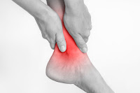
Sport's Dreaded "High Ankle Sprain"

Connecting the two bones of the lower leg & ankle (the tibia and fibula) are four ligaments called the syndesmotic ligaments. A high ankle sprain occurs with damage to one or more of these ligaments. This injury gets its common name because these ligaments are further up the leg than those damaged in other ankle sprain injuries. 

Causes

  • Injuries of the syndesmosis are commonly associated with an ankle fracture where one or more of these ligaments is partially or completely ruptured (torn).
  • Without fractures, sprains can occur when the foot is in an up position relative to the ankle and the leg rotates externally (outward). High ankle sprains may often follow, especially in cases where continued rotation causes a complete tear of the ligaments.
  • Being hit or kicked in the outside of the lower leg in soccer, football or other sports may cause injury to the syndesmosis. In these cases, other injuries along the inside of the ankle must be ruled out before diagnosing a high ankle sprain.


Signs & Symptoms

  • For acute injuries, swelling and pinpoint tenderness along the syndesmosis is often seen. Squeezing the lower leg muscles from side to side (the "squeeze test") may also cause pain in the area.
  • Moving the foot up and rotating it to the outside will also cause pain along the syndesmosis (low ankle sprains cause pain when the foot points down and in, contrasting the difference between the two).
  • May be associated with swelling along the inside or outside of the ankle when other injuries are suspected.
  • Difficulty bearing weight for moderate to severe injuries is extremely common. For more mild injuries, the patient may be able to bear weight.


X-Ray


X-rays of the ankle are necessary to rule out a separation of the tibia and fibula, especially in injuries that cause disruption of all four ligaments. However, x-rays may not be able to completely evaluate an injury to the syndesmosis when some of the four ligaments are intact. X-rays will also help to rule out any bone injury that may be associated with injuries to the syndesmosis. Stress testing may be performed under fluoroscopy, which allows us to move the foot in a certain position to try to produce a separation between the two ankle bones.


MRI


MRIs are more definitive for syndesmotic injuries. Ruptures of one or more of the ligaments are easily seen on the axial images. MRIs are also able to evaluate any abnormal position of the syndesmosis, which could contribute to long-term problems.


CT Scans


CT Scans can be used to evaluate the position of the tibia relative to the fibula. They are most often used in cases of this injury.


Treatment


For isolated injuries without a separation of the bones, conservative care is performed in almost all cases. This may include a short period of immobilization with or without weight-bearing activity. Physical therapy follows to encourage range of motion, strength and stability gains with a return to sports and activities. Ligaments heal reliably without any long-term problems if the entire syndesmosis has not been torn those these injuries typically take longer to heal than do low ankle sprains.
Example of an ankle sprain where complete rupture of
the syndesmosis resulted in surgery.

Complete ruptures of the syndesmosis, where the tibia and fibula are shown to be separated need to be addressed surgically. Fractures to the bones must always be ruled out first. Procedures are perfomed on an outpatient basis under a twilight or general anesthetic. One or two screws are placed from the fibula into the tibia to reduce the bone separation and allow healing of the ligaments. Immobilization in a boot or cast may be necessary for 4-6 weeks. However, using crutches to minimize weight-bearing activity is necessary for up to twelve weeks. Screws may or may not be removed for three months. We often replace the screws with large caliber sutures that maintain the reduction of the syndesmosis and allow normal motion of the ankle joint. At times these large sutures (tightropes) may be used in lieu of screws to prevent us from doing secondary surgical procedures. The degree of injury often dictates whether we use screws or suture buttons to repair the syndesmosis.


Prognosis


  • For mild injuries to the syndesmosis, conservative care produces an excellent long-term outcome. A return to sports within 6-8 weeks usually follows conservative care. Occasionally an impingement may develop in the ankle, necessitating a small cortisone shot to restore better motion to the ankle. A full recovery is expected in most cases.
  • When surgery is performed for moderate to severe injuries, the prognosis is excellent following a screw or suture button reduction of the syndesmosis. Return to sports may take 4-6 months. It is expected that our patients will continue to improve for 12-18 months following any surgical procedure. It is imperative to reduce the syndesmosis accurately to prevent any malalignment.


Use our online scheduler to book your appointment! If you have a busy schedule during the week, we are open Saturdays by appointment only in our recently redone Plantation office.


Click here to book your appointment and get back to your best!

Click here for pre- and post-surgery x-ray imaging and more information from Dr. Robert Sheinberg, DPM.

Tuesday, June 11, 2019

How to Get Rid of Plantar Fasciitis


How can I get rid of plantar fasciitis?
Do you suffer from Heel Pain?
PROBLEM:
  • An inflammation of the main ligament in the arch where it attaches to the heel bone and supports the foot.
CAUSES:
  • Excessive activity over a short period of time. 
  • Flat or high arched feet.
  • Tight muscles, especially the calf and hamstrings.
  • Poor shoe gear or walking barefoot for prolonged periods of time.
SYMPTOMS:
  • Pain first thing in the morning when getting out of bed and putting the foot down to the ground.  With walking the pain usually diminishes.
  • Pain after sitting for a prolonged period of time or getting out of a car and starting to walk.
  • Occasionally burning, numbness, shooting or tingling into the heel.
  • Extreme tenderness to touch the heel or arch region.
  • Commonly associated with lower back pain.
TREATMENT:
  • Avoid going barefooted and good supportive shoe wear.
  • Anti-inflammatory medications to reduce the inflammation.
  • Taping the foot to support it and give immediate relief.
  • Custom molded orthotic (shoe insert) to permanently support the foot and prevent reinjury.
  • Frequent stretching of the calf and hamstring muscles to improve overall flexibility.
  • Splinting the foot at night to stretch the muscle in the back of the leg.
  • Occasionally immobilization in a cast or a boot to completely rest the foot.
  • Shockwave therapy if pain persists.
  •  Surgery is a last resort to release a small portion of the ligament from the heel.

Friday, June 7, 2019

Crossover Digits on Your Foot

Predislocation Syndrome (Synovitis)


The Problem


Drifting of the second toe towards the big toe causing pain, deformity and early development of an overlapping second toe over the big toe. The second toe begins to curl as well.

Causes


  • Trauma to the second toe joint causing inflammation to that region.
  • Inflammation of the nerves on the ball of the foot (neuroma), weakening the joint.
  • Long second toe or metatarsal, causing a hammertoe and excess stress on the ball of the foot.
  • Weakness or tearing of the outer joint ligaments holding the second toe straight.
  • Stretching or tearing on the ligament on the ball of the foot called the "plantar plate".
  • Pressure on the second toe from a bunion deformity causing the toe to drift.
  • Pressure on the second toe due to a crooked big toe (hallux deformity).

Signs & Symptoms


  • Visible space between second and third toe while bearing weight. The third toe may also drift inward.
  • Pain to the second toe joint (both top and bottom) that increases with walking.
  • Difficulty walking barefoot and inability to wear heels.
  • Drifting of the second toe towards or over the first toe (overlapping toe).
  • Hammertoe of the second toe.
  • Neuritis (inflamed nerve causing numbness, burning and/or tingling).
  • Associated with bunion deformity and/or crooked big toe.
  • Swelling of the second toe and/or ball of the foot.
  • Callus (hard skin) on the ball of the foot because of metatarsal overload.

Treatment Options


  • Anti-inflammatories to decrease inflammation in the area.
  • Stiff-sole shoes to prevent toe bending.
  • Avoiding heels
  • When there is difficulty walking, temporary immobilization in a boot can eliminate pain.
  • Taping of the toes to lessen pain and deformity.
  • A small, carefully-directed cortisone injection can help in the case of severe pain. A boot will be necessary with it.
  • Surgery to realign the joint and help achieve a fully recovery. Surgery may include:
    • Ligament repair
    • Shortening the metatarsal (weil osteotomy)
    • Fusion of the second toe
    • Removal of inflamed tissue (synovectomy)
A pin may be necessary in the toe for up to six weeks. It would also be necessary to walk in a boot. A correction of a bunion at the same time may be necessary as it can be the underlying cause of the deformity.

Prognosis


Excellent with regard to eliminating the pain and lessening the deformity. The earlier the treatment, the better the long-term prognosis.


Use our online scheduler to book your appointment! If you have a busy schedule during the week, we are open Saturdays by appointment only in our recently redone Plantation office.


Click here to book your appointment and get back to your best!


Monday, June 3, 2019

Why Does My Child Walk On His/Her Toes?


Treatment:
1. In patients with mild tightness of the calf muscles, a home exercise program in patients with mild tightness that encourages proper calves (gastroc stretching) may be all that is necessary to achieve loosening of the calf muscles so that the motion goes back to the ankle instead of the foot.
2. NIGHT SPLINTS are often utilized to keep the foot at a 90-degree angle while the patient is sleeping. It may be utilized in mild cases of gastric solely (calf muscle) tightness.
3. PHYSICAL THERAPY may provide some limited benefits in patients with mild cases of muscle tightness. If there is a significant amount of tightness to the calf muscle, physical therapy will be of very limited benefit.
4. SERIAL CASTING is a procedure performed to loosen the tight calf muscles. The procedure is usually performed one leg at a time so that the patient can walk as much as possible while in the cast. The procedure is usually performed by placing a hard fiberglass cast that is not removable. It is applied from an area just below the knee to the base of the toes. While putting the cast on the foot is manipulated into its neutral position to avoid a collapsing arch. All the stress goes to the ankle and calf muscle during walking and the muscle undergoes a stretching process. The more the patient walks, the more the muscle stretches. This takes place because during gait the knee expands and the muscle stretches because the calf muscle attaches above and behind the knee. Casts are usually changed every two weeks until good mobility is present in the ankle and the calf muscle is no longer tight. In mild cases this may take 2 weeks and in more advanced cases it may take 4-8 weeks. Once one extremity is finished in the casting process a custom molded orthotic is made for the foot. The other leg is then casted in similar fashion. Casting both legs at the same time does not allow the patient to walk with knee extension; therefore, IT DOES NOT WORK. 
THE KEY WITH SERIAL CASTING IS TO HAVE THE PATIENT WALK AS MUCH AS POSSIBLE AND RESUME ALMOST ALL ACTIVITIES EXCEPT RUNNING AND JUMPING. When the casting is complete, night splints are used for a period of time to maintain the correction.
5. ACHILLES TENDON LENGTHENING SURGERY may be necessary in cases in which the calf muscle is exceptionally tight and the arch is completely collapsed. One of the most important procedures that we perform in our practice is to lengthen the calf muscle or Achilles tendon when serial casting or other measures have failed. The Achilles tendon lengthening procedure is done on an outpatient basis under a twilight or general anesthetic. Three small incisions in the back of the Achilles tendon are made, all 1/8” in length. The incisions are separated by 2.5 to 3 cm. One stitch is placed in each one of the incisions. A cast is applied postoperatively and the patient is non weightbearing for 6-8 weeks. Night splints are often utilized once the cast is removed and the patient begins physical therapy. This procedure is usually done with other procedures at the same time such as an “arthroeresis” or implant placed in the side of the foot to correct a flat foot. Full recovery can take three or more months. 

👣 Foot and ankle injury prevention and treatment should be easy ... with South Florida Institute of Sports Medicine, it is!

✅ South Florida trusts us to get them back to their best.


Make your appointment online at www.SouthFloridaSportsMedicine.com

Friday, May 31, 2019

My Child Is Complaining of Heel Pain

What Is Causing It and How Can I Help Them?

By Dr. John Goodner

Dr. Goodner is a Board Qualified
Reconstructive Foot & Ankle Surgeon
specializing in sports medicine foot and
ankle injuries, arthroscopy, trauma &
deformities in patients of all ages.
Painful inflammation to the growth plate (calcaneal apophysis) in the back of the heel is know as Sever's Disease. This ailment is most common in young soccer, baseball and basketball players due to the cleat patterns on shoes for these sports along with the specific demands these sports place on the heel region.

Why is my child having heel pain?


This type of heel pain typically occurs after overactivity in children between the ages of eight and fourteen. Tight muscles in their lower extremities, especially the calves and hamstrings, are usually to blame for the pain's onset. 

Many children who experience heel pain like this have associated flatfeet as well. Growth spurts can aggravate the condition further due to your child's body trying to adjust to the increased tension from already tight muscles on the calcaneal apophysis. Poor shoes or walking barefoot on hard surfaces can aggravate the heel's growth plates as well.

How do I know if my child has Calcaneal Apophysitis (Sever's Disease)?


  • Your child will display discomfort in the heel bone at the end of an activity. 
  • You will typically see stiffness in their foot in the morning and pain while walking. 
  • Their pain will be alleviated by rest and a decrease in activity.
  • Stiffness and pain will usually present after a child is done with activity, sits for a period of time and then tries to get up and walk.
  • Tenderness to the back, sides and bottom of the heel from touch or grasping of the heel.
  • You may notice your child walking on the ball of their foot to avoid touching their heel to the ground.
  • Discoloration and swelling likely won't be seen.
  • Limping at the end of activity that persists as the condition worsens is another sign.

How can my child's pain get better?


There is always a need to modify or avoid activities that aggravate the injury and avoid going barefooted. Taping the foot to decrease pulling on the plantar fascia is a good relief option. Proper shoes along with heel lifts can decrease pressure and tension on the heel.

Night splinting to stretch tight calf muscles and arch ligaments can maintain flexibility gained from calf stretching. Custom molded orthotics are necessary to support the foot and help to restore proper alignment in cases where flat feet contribute to the heel pain.

Physical therapy to improve flexibility and diminish inflammation of the growth plate may be necessary if symptoms persist after these home care options and activity modification do not work. Occasionally, immobilization in a cast or brace to completely rest the heel and allow healing may be necessary. 

Prognosis is excellent in most cases when a treatment plan is followed.


Use our online scheduler to book your appointment! If you have a busy schedule during the week, we are open Saturdays by appointment only in our recently redone Plantation office.


Click here to book your appointment and get your child back to their best!


Tuesday, May 28, 2019

5 Things you should know about: Running and your feet


Attention Runners! Here are some tips to avoid injuries.

Foot, Ankle and Leg injury? We offer same day appointments!

👣 Foot and ankle injury prevention and treatment should be easy ... with South Florida Institute of Sports Medicine, it is!

South Florida trusts us to get them back to their best.

Make your appointment online at www.SouthFloridaSportsMedicine.com

Monday, May 27, 2019

Ankle Arthroscopy Might Be the Solution You Need

Why does my ankle hurt??


What might have caused it?


Foot and ankle injuries are very common in sports but are also caused by regular activities in your daily life. There are many structures, including bones, ligaments, cartilage and joint lining around the ankle that may be damaged as a result of an injury.

Injuries can happen from a single traumatic event, from overuse, or from a previously untreated injury that manifests itself later in life. Joints like the ankle are designed to give our bodies freedom of movement, and any injury to them may alter your lifestyle.


Ankle Injury Symptoms


  • Injuries may cause stiffness and pain in the foot or ankle joint, especially when you first get out of bed in the morning and start to walk around. The stiffness/pain will be present for a period of time proportional to the degree of injury in the joint.
  • With weight-bearing, the joint usually loosens and the person is usually able to walk about or even run. Stiffness and pain will redevelop if the person sits for a period of time then tries to get back up and walk again.
  • As the day continues the foot or ankle may begin to ache, especially with excessive weight bearing.
  • Swelling may be present over the injured area.
  • Instability may develop if the ligaments have been injured, causing the foot or ankle to constantly twist.

Ankle Injury Treatment


  • When these injuries are unresponsive to conservative treatment (including orthotics, oral medication or injections, or rehabilitation) arthroscopic surgery may be necessary to remove the damaged tissue in the foot and ankle.


Ankle Arthroscopy Procedure


A miniature camera (between 1/8 and 1/6 inch) is placed into the injured joint and begins transmission to a television screen.The joint is visualized and small instruments, including a laser, are introduced into the affected area to clean out and repair the damage. 

Often, surgery includes removal of bone and/or cartilage fragments. Following that, we suction debris and smooth rough surfaces, often allowing for a joint to resume its normal gliding motion.

If cartilage is torn, loose or lifted, it is removed and small holes (microfractures) are placed in the bone to allow a regrowth of the cartilage. Although not the original cartilage (hyaline) the resurfacing with fibrocartilage is beneficial. Biocartilage implantation may also be performed to transplant cadaver cartilage and provide scaffolding for cartilage regrowth.

Conditions Treated by Ankle Arthroscopy


Advantages of Ankle Arthroscopy


  • Minimally invasive, with a faster recovery time
  • Less pain and swelling
  • Avoidance of large incisions and scars
  • Lower risk of infection
  • Outpatient procedure
  • Quicker return to regular activities and sports

When conservative treatments fail to alleviate pain and deformity of a joint, arthroscopic surgery should be considered. The surgical procedure is especially helpful for a fracture in the ankle join even after the ankle joint has been fixed surgically. Cartilage in the ankle joint may have been damaged during injury and cause chronic stiffness and pain that may be mild but never completely goes away.

Patients often think this is a condition they can live with, however, once treated arthroscopically a full return to activity may be possible. It's always the best treatment option for athletes and dancers.

Use our online scheduler to book your appointment! If you have a busy schedule during the week, we are open Saturdays by appointment only in our recently redone Plantation office.


Click here to book your appointment and get yourself back to your best!